
Reda Fadel1*, Fatima Zahra Lkharrat2, Abdelilah Arioua1, Oumayma Guennoun1, Zakaria Toubi1, Nizar Bouardi2, Mustapha Maaroufi2, Dounia Kamal1, Mohamed Nouredine El Amine El Alami1
1Service de stomatologie et de chirurgie maxillo-faciale, Hôpital OMAR IDRISSI, CHU HASSAN II, USMBA, FEZ
2Service de radiologie, Hôpital des spécialités, CHU HASSAN II, USMBA, FEZ
*Corresponding authors: Reda Fadel, Veltischev Research and Clinical Institute for Pediatrics and Рediatric surgery of the Pirogov Russian National Research Medical University, Moscow, Russia.
Received Date: August 16, 2023
Accepted Date: August 24, 2023
Published Date: August 30, 2023
Citation: Reda Fadel, Fatima Zahra Lkharrat, Abdelilah Arioua, Oumayma Guennoun . Zakaria Toubi (2023). “A Nasal Floor Ectopic Tooth: An Unusual Location from Diagnosis to Management.”. Clinical Research and Clinical Case Reports, 4(1); DOI: http;//doi.org/08.2023/1.1067.
Copyright: © 2023 Reda Fadel. This is an open access article distributed under the Creative Commons Attribution License, which permits unrestricted use, distribution, and reproduction in any medium, provided the original work is properly cited.
Ectopic tooth eruption is an uncommon dental anomaly where a tooth erupts or migrates to an abnormal location in the oral cavity or adjacent structures. We present the case of a 25 year old male who presented to our department with an ectopic tooth in the nasal floor, and share his challenging surgical management step by step. We also comment the use of PRF in the extraction site for guided bone regeneration that appears to be a promising adjunct to enhance bone healing and tissue regeneration.
Introduction:
Ectopic tooth eruption is an uncommon dental anomaly where a tooth erupts or migrates to an abnormal location in the oral cavity or adjacent structures. Among the various locations of ectopic tooth eruption, one rare and challenging site is the nasal floor. The presence of a tooth in the nasal floor can lead to significant complications, including recurrent sinus infections, nasal obstruction, and pain. Surgical extraction is often required to alleviate symptoms and prevent further complications. In recent years, Platelet-Rich Fibrin (PRF) has emerged as a promising adjunct in oral surgery, promoting tissue regeneration and enhancing wound healing. In this case report, we present a unique case of an ectopic tooth found in the nasal floor and describe the successful extraction procedure with the application of PRF for guided bone regeneration. Additionally we shed light on the management of such rare and challenging cases. [1, 2, 5]
Case report:
A 25 year old male presented to our department with a chief complain of a left maxillary swelling progressively increasing in size over the past three years. The patient associated the swelling to his decayed teeth and proceeded to their extraction. Upon examination the patient lacked his left maxillary incisors ( 21 and 22 ), the attached mucosa was sound, the swelling was of a bony nature and showed from the palatal side as well. No history of trauma, no bleeding, and no pain were reported. The rhinoscopy was normal, and the patient reported no nasal symptoms.
An orthopantomogram was performed and showed a left maxillary heterogenous lesion with a clear opaque component overlaying the nasal floor. A CT enhanced scan was then performed and was in favor of an ectopic nasal floor tooth with an enlarged peri coronary cyst.
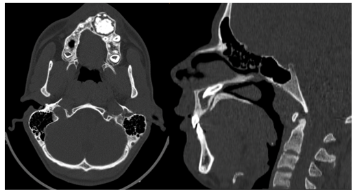
Figure1: CT scan imaging showing on the left an axial view the heterogenous filling of the cyst near the alveolar ridge, And on the right a sagittal view of the entrapped tooth in the nasal floor.
After the patient’s consent, surgery was performed, consisting in a cyst enucleation and a teeth extraction. First a crestal incision was performed prolonged by two vestibular counter-incisions allowing the raising of the mucoperiosteal flap. A bony alveolar window was then drilled with a rounded diamond bur. A heterogenous cyst was discovered and enucleated.
We then reached and extracted the ectopic tooth that was lying beneath the nasal septum. The nasal floor was not breached during extraction, and the bleeding was easily controlled. A 20 cc blood sample was drawn from the patient, and centrifuged in a codified fashion to obtain PRF. Once the PRF harvested, it was carefully placed in residual cavity in order to facilitate bone regeneration. The wound was closed with VICRYL 3-0 interrupted sutures. And post operative care was uneventful.
Discussion:
The presence of an ectopic tooth in the nasal floor is a rare anatomical finding and can cause significant discomfort and complications for the patient. In this case, the ectopic tooth was discovered incidentally during routine radiographic evaluation for a suspected sinus pathology. The accurate diagnosis and appropriate treatment of such cases are crucial to prevent potential complications such as recurrent sinus infections, pain, and nasal obstruction. [3,4] The extraction of an ectopic tooth from the nasal floor is a challenging surgical procedure, requiring meticulous planning and execution. In our case, we opted for a minimally invasive approach to minimize the risk of damaging nearby structures. The use of PRF for guided bone regeneration was a novel and promising adjunct to enhance bone healing and promote tissue regeneration at the extraction site. PRF is an autologous platelet concentrate rich in growth factors, which plays a significant role in tissue repair and regeneration. Its application in dentistry
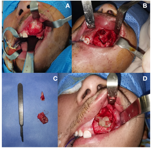
Figure2: A : Surgical approach and the mucoperiosteal flap raised. B : The extraction cavity and the residual tooth socket in the nasal floor after surgical removal. C : The tooth and the cyst after extraction. D : The residual cavity filled with PRF for guided bone regeneration before closure.
especially in oral surgery, has shown promising outcomes for bone augmentation and wound healing. In this case, we utilized PRF to facilitate guided bone regeneration at the ectopic tooth extraction site, aiming to restore bone volume and promote proper wound healing.The post- operative follow-up of the patient revealed satisfactory healing, with minimal complications. The use of PRF appeared to contribute to the overall success of the procedure, as evidenced by the enhanced bone formation and uneventful healing process.While the use of PRF for guided bone regeneration in ectopic tooth extraction is a promising approach, further studies with larger sample sizes and longer follow-up periods are needed to validate its efficacy and long-term outcomes. Additionally, comparative studies comparing different bone regeneration techniques would be valuable to establish the superiority of PRF in such cases. [5,6]
Conclusion:
The extraction of an ectopic tooth from the nasal floor is a rare and challenging procedure. The application of PRF for guided bone regeneration appears to be a promising adjunct to enhance bone healing and tissue regeneration at the extraction site. However, further research is warranted to fully assess the long-term benefits and efficacy of PRF in such cases. Early diagnosis and appropriate treatment are crucial to prevent potential complications and improve patient outcomes in ectopic tooth cases.
Acknowledgements:
No conflict of interest, nor funding is to be declared.

Open Access By Aditum Open Access Journals id licensed under Creative Commons Attribution 4.0 International License. Based On a Work at aditum.org